https://www.youtube.com/watch?v=UMTDmP81mG4
To license this video for patient education, content marketing or broadcast, visit: https://healthcare.nucleusmedicalmedia.com/contact-nucleus Reference: ANH12082
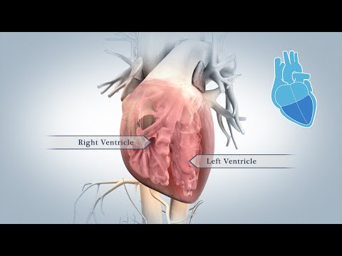
This 3D medical animation depicts the anatomy of the heart. Your heart is an extraordinary machine. During your lifetime, your heart’s muscular walls pump blood into blood vessels branching throughout your body.
Your heart has four chambers. Two upper chambers, called the left and right atria, and two lower chambers, called the left and right ventricles, contract in a steady rhythm known as your heartbeat. During a normal heartbeat, blood from your tissues and lungs flows into your atria, then into your ventricles.
Walls inside your heart, called the interatrial and interventricular septa, help keep the blood on left and right sides from mixing. Two valves sit like doors between your atria and ventricles to prevent blood from flowing backward into your atria. The tricuspid valve opens into your right ventricle, and the bicuspid valve, also known as the mitral valve, opens into your left ventricle.
Strong, thin tissues called chordae tendinae hold your valves in place during the forceful contractions of your ventricles. Blood leaving the ventricles passes through another set of valves: the pulmonary valve, between your right ventricle and pulmonary trunk, and the aortic valve, connecting your left ventricle and aorta.
In order to pump blood more efficiently, your heart muscle, called myocardium, is arranged in a unique pattern. Three layers of myocardium wrap around the lower part of your heart. They twist and tighten in different directions to push blood through your heart. When special cells, called pacemaker cells, generate electrical signals inside your heart, the heart muscle cells, called myocytes, contract as a group.
Your heart is divided into left and right halves, which work together like a dual pump. On the right side of your heart, deoxygenated blood from your body’s tissues flows through large veins, called the superior and inferior vena cava, into your right atrium. Next, the blood moves into your right ventricle, which contracts and sends blood out of your heart to your lungs to gather oxygen and get rid of carbon dioxide.
On the left side of your heart, oxygen-rich blood from your lungs flows through your pulmonary veins into your left atrium. The blood then moves into your left ventricle, which contracts and sends blood out of your heart through the aorta to feed your cells and tissues.
The first branches off your aorta are the coronary arteries, which supply your heart muscle with oxygen and nutrients. At the top of your aorta, arteries branch off to carry blood to your head and arms. Arteries branching from the middle and lower parts of your aorta supply blood to the rest of your body.
Your heart beats an average of sixty to one hundred beats per minute. In that one minute, your heart pumps about five quarts of blood through your arteries, delivering a steady stream of oxygen and nutrients all over your body.
#HeartAnatomy #CardiacAnatomy #Heart


